A white spot or patch on your tongue is often harmless, but the most useful clue is whether it wipes off. If it rubs off and leaves a red or slightly bleeding surface, it is more likely oral thrush, a common yeast infection. If it stays put, it may be a thickened patch from rubbing or something that needs a dentist’s eyes. Any spot that lasts about two weeks or comes with red-flag symptoms should be checked.
If you have trouble breathing, swelling in the mouth or throat, or severe pain, do not wait for a two-week check. Seek same-day care.
Wipeable or Not? A Simple First Step
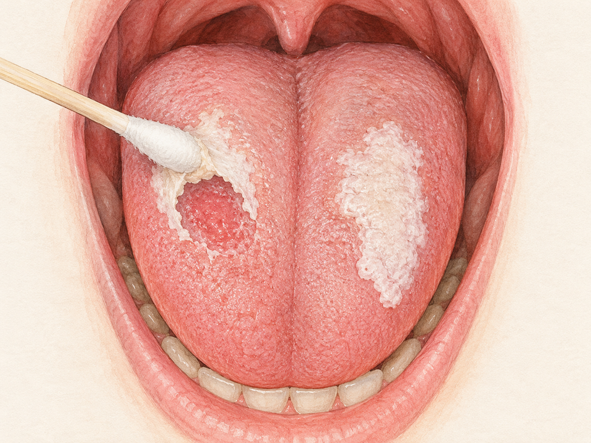
Dentists often start with the same question: does the white area rub off? You can gently wipe the spot with a damp cotton swab or gauze. Do not scrub.
- If it wipes away and leaves a red or bleeding surface, oral thrush is more likely. Thrush is a yeast infection in the mouth. It can look like creamy white patches, sometimes described as curdled milk.
- If it does not wipe off, the white area may be a thickened patch of surface cells. Two examples are leukoplakia, a persistent white patch, and frictional keratosis, a rough white patch from rubbing against teeth or dentures.
This wipe test is only a clue, not a diagnosis. A clinical decision tree for oral white lesions uses this same step to narrow down possibilities. It helps you and your dentist think through what might be going on.
Common Causes That Usually Aren’t Serious
Several everyday issues can cause a white spot or coating on the tongue.
- Oral thrush: Creamy white patches that may wipe off. They can leave redness or slight bleeding. Thrush is more common after antibiotics, inhaled steroid use, or when the immune system is low. It usually improves with antifungal treatment, but underlying triggers may need attention.
- Geographic tongue: Smooth red patches with white or gray borders that look like a map. It can come and go and is usually harmless.
- Lie bumps: Tiny, sometimes tender bumps that can look white or pink. They usually fade within a few days.
- Friction or denture irritation: A white patch where a tooth, sharp edge, or denture rubs. It often improves once the source of rubbing is removed, according to guidance on oral white lesions.
- Minor burn: A white or sore area after hot food or drink. It should heal in about one to two weeks.
- Dry mouth or dehydration coating: A pasty white coating on the tongue, often thicker toward the back. Drinking fluids and gently cleaning the tongue may help.
Most of these improve with simple changes or treatment. But if the spot lasts beyond about two weeks or gets worse, have it checked.
Causes That Need a Dentist’s Eyes
Some white changes need a professional look.
- Leukoplakia: A persistent white patch that does not wipe off. It can be caused by tobacco, friction, or unknown reasons. Some leukoplakia patches are harmless, while others carry a small risk of becoming cancer. A dentist cannot always tell which is which just by looking, so persistent patches are usually evaluated.
- Oral lichen planus: A long-term immune-related condition that can create white, lacy lines or patches. It may come and go, and it sometimes needs treatment or regular monitoring. It is not cancer, but some forms need follow-up.
- Persistent ulcer or sore: A sore that looks white or has a white coating and does not heal could be from repeated trauma, infection, or a more serious problem. It needs professional evaluation.
The two-week rule is a good guide: if a white spot or patch is still there after about two weeks, see a dentist. This does not mean you should panic. It means a professional should take a look. Living evidence-informed guidance says biopsy or referral should be considered when a mouth abnormality persists for 10 to 14 days without a clear diagnosis.
You cannot diagnose these conditions from a photo or a symptom list. Many require a visual exam and sometimes a tissue sample.
Red Flags That Mean Don’t Wait
Some signs are more urgent than the usual two-week watch.
- A firm bump in the mouth or on the tongue
- Bleeding from the white area without a clear cause
- A white or red area that is clearly getting bigger
- Pain when swallowing
- Ear or jaw pain on the same side
- A new or changing lump in the neck
- Fever or trouble breathing
These can point to a deeper infection, a problem outside the mouth, or, in rare cases, a cancer. They deserve same-day care, not a two-week wait. Guidance on early detection of head and neck cancers highlights similar red-flag symptoms that should trigger prompt referral.
Simple Things to Try While You Wait
These steps may help while you wait for an appointment. They are not a treatment.
- Drink plenty of water to keep your mouth moist.
- Rinse gently with warm salt water if it feels soothing.
- Gently clean your tongue with a soft brush or scraper only if it does not irritate the area.
- Remove obvious irritants such as tobacco, harsh mouthwashes, or an ill-fitting denture if you can do so safely.
- Avoid picking at the spot or scrubbing it hard.
- Do not use antifungal or steroid creams unless a clinician has diagnosed the cause. Using the wrong cream can mask symptoms or irritate the area.
Who to See First: Dentist or Doctor
A dentist is usually the right first stop for a white spot on the tongue, especially if it is painless and persistent. Dentists are trained to examine mouth tissue and can refer you for a biopsy if needed. Guidance on evaluating suspicious oral lesions describes a step-by-step approach that dentists use.
If you have whole-body symptoms, such as fever, weight loss, or pain when swallowing, or if the spot is linked to a known medical condition, your primary care doctor or urgent care may be the better first call. If you are not sure, a phone call to either office can help you decide.
What to Expect at the Appointment
Most visits for a white spot are straightforward and calm.
- History questions: The dentist will ask how long the spot has been there, whether it hurts, and about smoking, medicines, and general health.
- Visual exam: They will look carefully inside your mouth, including your tongue, cheeks, floor of the mouth, and roof.
- Gentle palpation: They may feel the area and nearby neck with gloved fingers to check texture and lumps.
- Possible swab: If a yeast infection is suspected, a swab may be taken from the surface.
- Possible biopsy: If the spot is unclear or concerning, a small tissue sample may be taken, or you may be referred to a specialist. A biopsy is the most reliable way to find out what the tissue is.
In most cases, the visit ends with reassurance, a treatable diagnosis like thrush, or a simple referral. Going in early is easier than waiting and worrying.
A white spot on your tongue is common and often minor, but you don’t need to guess. Use the wipe test as a clue, follow the two-week rule, and see a dentist if anything feels off.
تمت مراجعة هذا المقال من قِبَل مختص في صحة الفم لضمان دقته. يهدف المقال إلى تقديم معلومات تثقيفية عامة، ولا ينبغي استخدامه بديلاً عن المشورة الطبية أو التشخيص أو العلاج المخصص لحالتك. إذا كانت لديك أي أسئلة أو مخاوف تتعلق بصحة فمك، فيُرجى استشارة طبيب الأسنان أو الطبيب أو أي مقدم رعاية صحية مؤهل آخر.